Clinical Gross Anatomy of the Skin
|
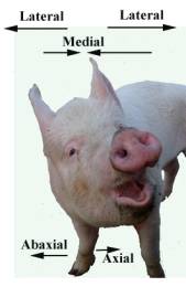
It is important to standardize the description and position of anatomical features. The following pictures provide illustrations of terms used to describe directions around the pig and major surface landmarks. |
||
|
Describing the general directions around the pig |
||
|
|
|
|
|
|
||
|
|
|
|
|
View of the pig neck |
The head view of the pig |
|


|
Surface landmarks relating to the sex of the pig |
|
|
The
major surface landmarks of the rear of the female pig: |
|
|
|
|
|
The hind view of a female gilt |
Detail of the vulva opened |
|
The entire male: |
The castrated male: |
|
|
|
|
Remember that the male also has the prepuce on the ventral surface. |
|
|
|
|
|
Lateral view of the mammary glands |
Detail of the teat with milk from the supplying mammae being expressed |


|
Specific detail of the
surface anatomy of the lower limb |
|
|
|
|
|
Detail of the dorsal surface of the front foot |
|
|
|
|
|
Plantar view of the foot |
|
|
|
|
|
Lateral view of the front foot |
Carpal glands – function unknown |


When clinically examining the pig, it is essential
to be able to visualize the position of the major organs.
The following illustrations reflect the major organs
onto the surface of the pig.
The white line represents the position of the last rib. The organs are similarly colored in each drawing. The organs are labeled in the right side illustration. Note the large size of the pig’s head to the rest of its body.
|
Left side Rectum Kidney Uterus Small intestine Stomach Liver Lung Heart |
|
|
|
|
Right side Kidney Rectum Uterus Large intestine Stomach Liver Lung Heart |
|
|
|
From the Dorsum |
|


|
Skeleton and organ layout superimposed on the image of the pig |
|
|
|
|
|
The commercial Large White/Yorkshire |
|
|
|
|
|
The Vietnamese Pot Belly |
|
|
|
|
|
Detail
of the major meat joints of the pig drawn on the skin of the pig |
|
|
|
|


