Clinical
Gross Anatomy of the Intestinal Tract
|
|
|||
|
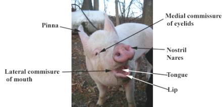
General view of the head of the pig |
|||
|
|
|
||
|
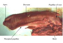
The tongue dorsal view |
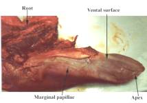
The tongue ventral view |
||
|
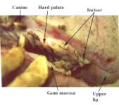
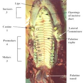
Detail of the gum margin and teeth |
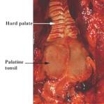
The hard and soft palate – ventral view |
||
|
Detail of the palatine tonsil |
|||
|
|
|
||
|
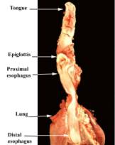
The oesophagus opened |
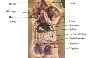
General view of the pig, ventral body wall removed |
||
|
|
|
||
|
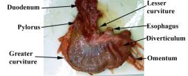
The parietal surface of the stomach |
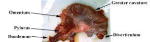
The visceral surface of the stomach |
||
|
|
|||
|
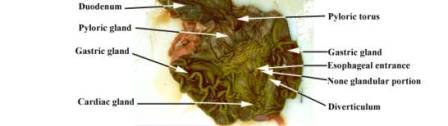
The mucosal surface of the stomach, the organ opened along the greater curvature |
|||
|
|
|
||
|
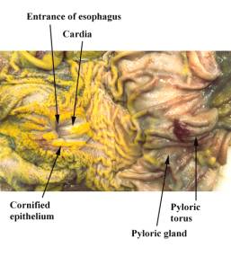
Detail of the oesophageal opening |
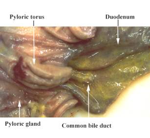
Detail of the pyloric sphincter opened |
||
|
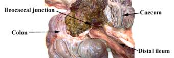
The ileocaecal junction |
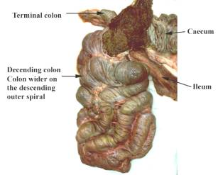
The spiral colon of a pig |
||
|
The ileocaecal ligament, useful in locating the ileocaecal junction |
|||
|
|
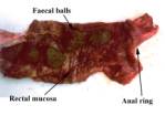
The rectum opened |
||
|
|
|
||
|
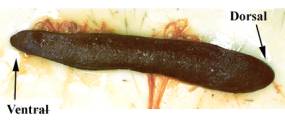
The spleen parietal surface |
The spleen visceral surface |
||
|
|
|
||
|
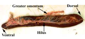
The liver parietal surface |
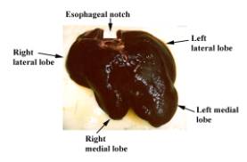
The liver visceral surface |
||
|
|
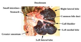
The liver, visceral surface showing the common bile duct. |
||
|
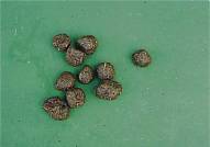
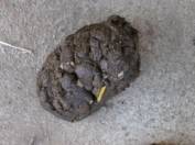
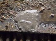
Feaces: It is always important to note the consistency and colour of feacal pellets: |
|||
|
|
|
|
|
|
Constipated |
Normal |
Diarrhoea |
|