Respiratory
defense mechanisms
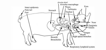
|
Organ |
Example defense |
Defense interfered with |
|
|
|
|
Disease/disorder |
Environmental |
|
Eyes |
Vision |
Chlamydia Conjunctivitis |
Dust Ammonia |
|
Tongue |
Taste |
|
Food -
palatability |
|
Mouth/pharynx |
Amylase Sneezing |
Aujeszky’s Disease Actinobacillus pleuropneumoniae Haemophilus parasuis Streptococcus
suis et al., Mycoplasma hyorhinis Pasteurella multocida E. coli PCV2 Other mycoplasma’s |
|
|
Tonsils |
Normal microbiota Lymphoid
tissue Rapid healing Washing with
liquids |
Mycotoxins |
|
|
Epiglottis/larynx |
Size Separates
swallowing and breathing |
|
|
|
Nose |
Small entrance |
|
|
|
Turbinates |
Size Mucocilary escalator down to throat High blood
volume High humidity Centrifugal
forces Smell Immune
response Natural microbiota Lysosymes and other antimicrobial agents in mucus. Sneezing |
Inclusion body
rhinitis Bordetella bronchiseptica Pasturella multocida toxins Post-weaning
sneezing Pasteurella multocidia Mycoplasma’s |
Ammonia Other poison
gases Dust Low water |
|
Trachea |
Length Corners/bends High air
velocity Centrifugal forces Mucocilary escalator up to throat Immune
response Lysosymes and other antimicrobial agents in mucus |
Mycoplasma hyopneumoniae Swine
Influenza |
Low water Ammonia Dust |
|
Right apical bronchus |
|||
|
Bronchus and bronchi |
|||
|
Lung general |
Ascaris suum Metastrongylus apri Classical Swine Fever Actinobacillus pleuropneumoniae Pasteurella multocidia Streptococci Archanobacterium pyogenes |
||
|
Alveolus |
Alveolar
macrophage Immune
response Alveolar fluid |
PRRSv |
Dust: 3.6-1 µm |
|
Pleura |
Immune
response - Fibrosis |
Glässer’s
Disease Actinobacillus
pleuropneumoniae Tail biting
and other vices |
Flooring |
|
Respiratory lymphoid tissues |
Immune
response |
PCV2 SIV |
Mycotoxins Low Vit E Colostrum
intake |
|
Stomach |
Acid Proteases Vomiting |
Gastric ulcer Hyostrongylus dentatum |
Low water Not eating Feed particle
size |
|
Intact skin epidermis |
Normal microbiota Continuous
surface |
Vice – tail
biting etc Damage to feet Fighting |
Rough floors Sharp edges Pig processing Injections |
Intestinal
defense mechanisms

|
Organ |
Example defense |
Defense interfered with |
|
|
|
|
Disease/disorder |
Environmental |
|
Eyes |
Vision Avoidance of
unusual food |
Chlamydia Conjunctivitis |
Food colouring – note pigs are colour
blind |
|
Nose |
Smell Avoidance of
unusual or unpalatable food |
|
Toxin smells |
|
Tongue |
Taste and feel
food |
|
Toxin taste Sharp objects |
|
Mouth/pharynx |
Teeth to break
up food particles Salivary
enzyme actions on sugars |
Aujeszky’s Disease Haemophilus parasuis Streptococcus
suis Mycoplasma hyorhinis PCV2 E. coli Salmonella Clostridia Teeth hygiene Tooth wear |
Stones Sham chewing |
|
Tonsils |
Normal microbiota Lymphoid
tissue Rapid healing |
Mycotoxins |
|
|
Epiglottis |
Size Separates
swallowing and breathing Coughing |
|
|
|
Stomach |
Acidic
environment Proteases Mucus Pyloric torus
protect stomach from alkaline |
Gastric ulcer Hyostrongylus dentantum |
Low water Not eating Feed particle
size |
|
Small intestine |
Sudden change
in pH Bile
production Digestive
enzymes Rapid cell
turn over (every 3 to 6 days) Mucus One way flow -
peristalsis Microbiota – Lactobacilli and Bacilli Large amounts
of diluting water Microaerophilic Lymphoid
tissue Epithelial
attachment Diluting
effect of water Genetic
attachment changes – F4 F18 Defensins Tight junction
epithelium Immune
response Diarrhoea |
Clostridium perfringens Clostridium difficile Enteric
Diarrhoea virus Escherichia coli Isospora suis Lawsonia intracellularis Salmonellosis TGE Trichuris suis Yersinia enterocolitica Strongyle/parasites PCV2 |
Myotoxins Vit E Low iron Low water
availability Poor colostrum
intake |
|
Large intestine |
Ileocaecal valve Diluting water
control One way flow -
peristalsis Change in O2
concentration Indigenous microbiota Diarrhoea |
Brachyspira hyodysenteriae Brachyspira pilosicoli Other Brachyspira |
|
|
Rectum/anus |
One way flow Defecation |
Rectal
prolapsed Salmonellosis |
Chilling and
piling Coughing |
|
Liver |
|
Clostridium novii |
|
|
Intact skin epidermis |
Normal microbiota Continuous
surface |
Vice – tail
biting etc Damage to feet Fighting |
Rough floors Sharp edges Pig processing Injections |
|
Organ |
Example defense |
Defense interfered
with |
|
|
|
|
Disease/disorder |
Environmental |
|
External genitalia |
Shape Flushing
sterile urine Normal microbiota |
Vulva biting |
Poor water
flow Other vices |
|
Urethra |
Male - length |
|
|
|
Bladder |
Transitional
layer Glycopolypeptide layer Cell loss Epithelial attachement, E.
coli F1, P Urination Flushing
sterile urine |
Actinobaculum suis Escherichia coli Streptococcus suis Crystaluria |
Water
restriction Feed toxins Floor hygiene Floor mobility
- stance |
|
Ureterovesical junction |
Length Colosure during urination Sterile urine |
||
|
Ureter |
Sterile urine |
||
|
Pelvis |
Sterile urine |
||
|
Pyamid shape – Duct of Belini |
Sterile urine |
||
|
Nephron |
Sterile urine |
Actinobaculum suis Leptospirosis |
|
|
Glomerulus |
Sterile urine Filter blood |
PDNS |
|
|
Vagina |
Robust immune defence Especially
under progesterone Normal microbiotia Flushing of
sterile urine |
AI
contamination Vaginitis with streptococci and Staphylococci et al., |
Water
restriction Floor hygiene |
|
Cervix |
Closure Flushing of
sterile urine Normal microbiota |
Cerviculitis with variety of environmental bacteria |
Water
restriction Floor hygiene |
|
Uterus |
Robust immune defence under progesterone |
Parvovirus Aujeszky’s Disease PRRSv Enterovirus JEV Streptococci Leptospirosis Brucella suis PCV2 Congenital
tremor virus Classical Swine
Fever |
Feed – mycotoxins Midwifery
practices Mating
practices |
|
Tubal junction/oviduct |
Controlled
flow Restricted
entry of semen |
|
|
|
Ovary |
|
Cystic ovaries |
“Stress”
issues |
|
Prepuce |
Normal microbiota High pH |
Preputial diverticulitis |
|
|
Vas Deferens |
Length Ejaculation |
|
|
|
Reproductive organs |
|
|
|
|
Testes |
Blood testes
barrier |
Brucella suis |
|